What Triggers Pain On The Heel To Surface

Overview
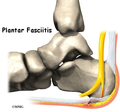
Plantar fasciitis is the most common cause of heel pain. Pain from plantar fasciitis is often most noticeable during the first few steps after getting out of bed in the morning. The plantar fascia is a thick band of tissue in the sole of the foot. Microtearing at the origin of the plantar fascia on the heel bone (calcaneus) can occur with repetitive loading. This microtearing leads to an inflammatory response (healing response) which produces the pain. Risk factors for plantar fasciitis include excessive standing, increased body weight, increasing age, a change in activity level, and a stiff calf muscle. Plantar fasciitis can be managed non-operatively in the vast majority of patients. The main components of an effective non-operative treatment program are: calf stretching with the knee straight, plantar fascia stretching, activity modification (to avoid precipitating activities), and comfort shoe wear.
Causes
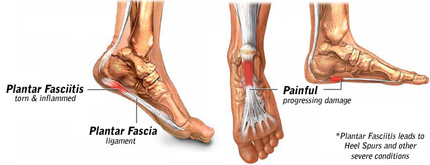
Training on improper, hard and/or irregular surfaces as well as excessive track work in spiked shoes, or steep hill running, can stress the plantar fascia past its limits of elasticity, leading to injury. Finally, failure in the early season to warm up gradually gives the athlete insufficient time for the structures of the foot to re-acclimate and return to a proper fitness level for intensive exercise. Such unprepared and repeated trauma causes microscopic tearing, which may only be detected once full-blown plantar fasciitis and accompanying pain and debilitation have resulted. If the level of damage to the plantar fascia is significant, an inflammatory reaction of the heel bone can produce spike-like projections of new bone, known as heel spurs. Indeed, plantar fasciitis has occasionally been refereed to as heel spur syndrome, though such spurs are not the cause of the initial pain but are instead a further symptom of the problem. While such spurs are sometimes painless, in other cases they cause pain or disability in the athlete, and surgical intervention to remove them may be required. A dull, intermittent pain in the heel is typical, sometimes progressing to a sharp, sustained discomfort. Commonly, pain is worse in the morning or after sitting, later decreasing as the patient begins walking, though standing or walking for long periods usually brings renewal of the pain.
Symptoms
The symptoms of plantar fasciitis include pain in the bottom of your foot, especially at the front or centre of the heel bone, pain that is worse when first rising in the morning (called "first-step pain"), when first standing up after any long period of sitting, or after increased levels of activity especially in non-supportive shoes. Seek medical advice about plantar fasciitis if you have heel pain or pain in the bottom of your foot, especially when you get up in the morning, that does not respond to treatment or if there is redness or bruising in the heel.
Diagnosis
Your doctor will ask you about the kind of pain you're having, when it occurs and how long you've had it. If you have pain in your heel when you stand up for the first time in the morning, you may have plantar fasciitis. Most people with plantar fasciitis say the pain is like a knife or a pin sticking into the bottom of the foot. After you've been standing for a while, the pain becomes more like a dull ache. If you sit down for any length of time, the sharp pain will come back when you stand up again.
Non Surgical Treatment
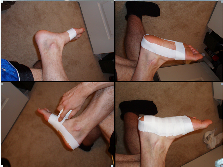
Conservative treatment is almost always successful, given enough time. Traditional treatment often includes, rest, NSAIDs, and new shoes or heel inserts. Some doctors also recommend avoiding walking bare-footed. This means you’d have to wear your shoes as soon as you wake up. Certain foot and calf exercises are often prescribed to slowly build strength in the ligaments and muscles that support the arch of the foot. While traditional treatment usually relieves pain, it can last from several months to 2 years before symptoms get better. On average, non-Airrosti patients tend to get better in about 9 months.

Surgical Treatment
The most dramatic therapy, used only in cases where pain is very severe, is surgery. The plantar fascia can be partially detached from the heel bone, but the arch of the foot is weakened and full function may be lost. Another surgery involves lengthening the calf muscle, a process called gastrocnemius recession. If you ignore the condition, you can develop chronic heel pain. This can change the way you walk and cause injury to your legs, knees, hips and back. Steroid injections and some other treatments can weaken the plantar fascia ligament and cause potential rupture of the ligament. Surgery carries the risks of bleeding, infection, and reactions to anesthesia. Plantar fascia detachment can also cause changes in your foot and nerve damage. Gastrocnemius resection can also cause nerve damage.
Prevention
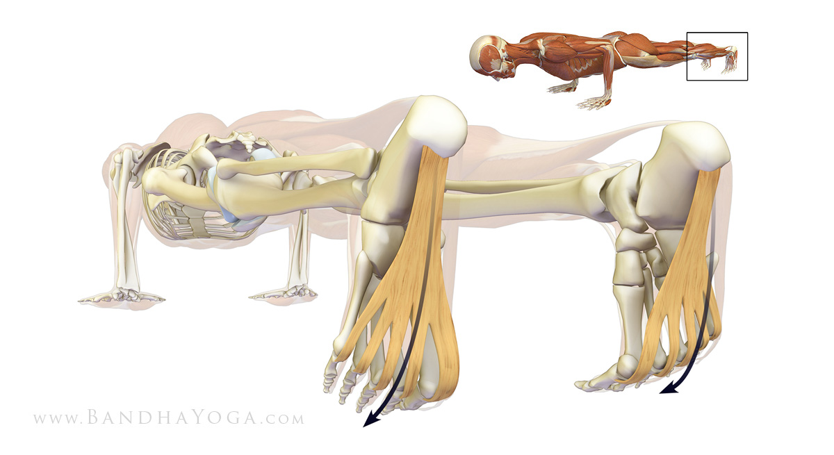
Stretching the plantar fascia and the calf muscle area can help to prevent inflammation. Slowly increasing the amount or intensity of athletic activities by graded progression can also help to prevent injury. Recommended Stretches: Taking a lunge position with the injured foot behind and keeping your heels flat on the floor, lean into a wall and bend the knees. A stretch should be felt in the sole and in the Achilles tendon area. Hold the stretch for 20-30 seconds. Also try this stretch with the back leg straight.
What Is Pain At The Heel
Overview
Plantar Fasciitis is a chronic pain in the heel that can just appear from nowhere but it is actually a long standing mechanical condition caused by prolonged stress on your foot. In most cases Plantar Fasciitis is a common, but very treatable, mechanical condition of the foot and responds positively to orthotics. A heel pain caused by prolonged stress on a ligament like structure in the arch that is very important in weigh-bearing activities. The tissue becomes damaged and needs to be helped to repair in order for the pain to go or subside to a manageable level. Orthotics for your feet can achieve this necessary healing for pain relief. It can be very painful, and even debilitating for sufferers.
Causes
Plantar fasciitis occurs when the thick band of tissue on the bottom of the foot is overstretched or overused. This can be painful and make walking more difficult. You are more likely to get plantar fasciitis if you Have foot arch problems (both flat feet and high arches), run long distances, downhill or on uneven surfaces, are obese or gain weight suddenly, have a tight Achilles tendon (the tendon connecting the calf muscles to the heel), wear shoes with poor arch support or soft soles. Plantar fasciitis is seen in both men and women. However, it most often affects active men ages 40 - 70. It is one of the most common orthopedic foot complaints. Plantar fasciitis was commonly thought to be caused by a heel spur. However, research has found that this is not the case. On x-ray, heel spurs are seen in people with and without plantar fasciitis.
Symptoms
Symptoms of the plantar fasciitis include a gradual onset of pain under the heel which may radiate into the foot. Tenderness is usually felt under and on the inside of the heel which is initially worse first in the morning but eases as the foot warms up only to return later in the day or after exercise. Stretching the plantar fascia may be painful.
Diagnosis
Plantar fasciitis is usually diagnosed by a health care provider after consideration of a person’s presenting history, risk factors, and clinical examination. Tenderness to palpation along the inner aspect of the heel bone on the sole of the foot may be elicited during the physical examination. The foot may have limited dorsiflexion due to tightness of the calf muscles or the Achilles tendon. Dorsiflexion of the foot may elicit the pain due to stretching of the plantar fascia with this motion. Diagnostic imaging studies are not usually needed to diagnose plantar fasciitis. However, in certain cases a physician may decide imaging studies (such as X-rays, diagnostic ultrasound or MRI) are warranted to rule out other serious causes of foot pain. Bilateral heel pain or heel pain in the context of a systemic illness may indicate a need for a more in-depth diagnostic investigation. Lateral view x-rays of the ankle are the recommended first-line imaging modality to assess for other causes of heel pain such as stress fractures or bone spur development. Plantar fascia aponeurosis thickening at the heel greater than 5 millimeters as demonstrated by ultrasound is consistent with a diagnosis of plantar fasciitis. An incidental finding associated with this condition is a heel spur, a small bony calcification on the calcaneus (heel bone), which can be found in up to 50% of those with plantar fasciitis. In such cases, it is the underlying plantar fasciitis that produces the heel pain, and not the spur itself. The condition is responsible for the creation of the spur though the clinical significance of heel spurs in plantar fasciitis remains unclear.
Non Surgical Treatment
A steroid (cortisone) injection is sometimes tried if your pain remains bad despite the above 'conservative' measures. It may relieve the pain in some people for several weeks but does not always cure the problem. It is not always successful and may be sore to have done. Steroids work by reducing inflammation. Sometimes two or three injections are tried over a period of weeks if the first is not successful. Steroid injections do carry some risks, including (rarely) tearing (rupture) of the plantar fascia. Extracorporeal shock-wave therapy. In extracorporeal shock-wave therapy, a machine is used to deliver high-energy sound waves through your skin to the painful area on your foot. It is not known exactly how it works, but it is thought that it might stimulate healing of your plantar fascia. One or more sessions of treatment may be needed. This procedure appears to be safe but it is uncertain how well it works. This is mostly because of a lack of large, well-designed clinical trials. You should have a full discussion with your doctor about the potential benefits and risks. In studies, most people who have had extracorporeal shock-wave therapy have little in the way of problems. However, possible problems that can occur include pain during treatment, skin reddening, and swelling of your foot or bruising. Another theoretical problem could include the condition getting worse because of rupture of your plantar fascia or damage to the tissues in your foot. More research into extracorporeal shock-wave therapy for plantar fasciitis is needed. Other treatments. Various studies and trials have been carried out looking at other possible treatments for plantar fasciitis. Such treatments include injection with botulinum toxin and treatment of the plantar fascia with radiotherapy. These treatments may not be widely available. Some people benefit from wearing a special splint overnight to keep their Achilles tendon and plantar fascia slightly stretched. The aim is to prevent the plantar fascia from tightening up overnight. In very difficult cases, sometimes a plaster cast or a removable walking brace is put on the lower leg. This provides rest, protection, cushioning and slight stretching of the plantar fascia and Achilles tendon. However, the evidence for the use of splint treatment of plantar fasciitis is limited.
Surgical Treatment
Surgery for plantar fasciitis can be very successful in the right patients. While there are potential complications, about 70-80% of patients will find relief after plantar fascia release surgery. This may not be perfect, but if plantar fasciitis has been slowing you down for a year or more, it may well be worth these potential risks of surgery. New surgical techniques allow surgery to release the plantar fascia to be performed through small incisions using a tiny camera to locate and cut the plantar fascia. This procedure is called an endoscopic plantar fascia release. Some surgeons are concerned that the endoscopic plantar fascia release procedure increases the risk of damage to the small nerves of the foot. While there is no definitive answer that this endoscopic plantar fascia release is better or worse than a traditional plantar fascia release, most surgeons still prefer the traditional approach.
What Is Pain In The Heel
Overview
Plantar fasciitis is the most common cause of heel pain. The plantar fascia is the flat band of tissue (ligament) that connects your heel bone to your toes. It supports the arch of your foot. If you strain your plantar fascia, it gets weak, swollen, and irritated (inflamed). Then your heel or the bottom of your foot hurts when you stand or walk. Plantar fasciitis is common in middle-aged people. It also occurs in younger people who are on their feet a lot, like athletes or soldiers. It can happen in one foot or both feet.
Causes
Plantar Fasciitis is caused by abnormal pronation of the foot. Contributing factors are obesity, weight gain, jobs that require a lot of walking or standing on hard surfaces, badly worn shoes with little support, and also inactivity. As a result of over-pronation, with every step the Plantar Fascia (band of tissue under the foot) is being stretched, resulting in inflammation, irritation and pain at the attachment of the fascia into the heel bone. In some cases the pain is felt under the foot, in the arch. Continuous pulling of the fascia at the heel bone, eventually may lead to the development of bony growth on the heel. This is called a heel spur. When you’re at rest, such as while sleeping, the Plantar Fascia tightens and shortens. When body weight is rapidly applied to the foot, the Fascia must stretch and quickly lengthen, causing micro-tears in the Fascia. As a result, the foot pain is more severe with your first steps in the morning, or after sitting for a long period. Plantar Fasciitis is more likely to happen if you suffer from over-pronation (flattening of the arch), you stand or walk on hard surfaces, for long periods, you are overweight or pregnant, you have tight calf muscles.
Symptoms
Plantar fasciitis has a few possible symptoms. The symptoms can occur suddenly or gradually. Not all of the symptoms must be present at once. The classic symptom of plantar fasciitis is pain around the heel with the first few steps out of bed or after resting for a considerable period of time. This pain fades away a few minutes after the feet warm up. This symptom is so common that it symbols the plantar fasciitis disorder. If you have it then probably you have plantar fasciitis. If you don’t suffer from morning pain then you might want to reconsider your diagnosis. Pain below the heel bone at the connection of the bone to the fascia. As the condition becomes more severe the pain can get more intense during the day without rest. Plantar fasciitis symptoms include pain while touching the inside of the heel or along the arch. Foot pain after you spend long periods of time standing on your feet. Pain when stretching the plantar fascia. Foot pain that worsens when climbing stairs or standing on the toes. Pain that feels as though you are walking on glass. Pain when you start to exercise that gets better as you warm up but returns after you stop.
Diagnosis
Plantar fasciitis is usually diagnosed by a health care provider after consideration of a person’s presenting history, risk factors, and clinical examination. Tenderness to palpation along the inner aspect of the heel bone on the sole of the foot may be elicited during the physical examination. The foot may have limited dorsiflexion due to tightness of the calf muscles or the Achilles tendon. Dorsiflexion of the foot may elicit the pain due to stretching of the plantar fascia with this motion. Diagnostic imaging studies are not usually needed to diagnose plantar fasciitis. However, in certain cases a physician may decide imaging studies (such as X-rays, diagnostic ultrasound or MRI) are warranted to rule out other serious causes of foot pain. Bilateral heel pain or heel pain in the context of a systemic illness may indicate a need for a more in-depth diagnostic investigation. Lateral view x-rays of the ankle are the recommended first-line imaging modality to assess for other causes of heel pain such as stress fractures or bone spur development. Plantar fascia aponeurosis thickening at the heel greater than 5 millimeters as demonstrated by ultrasound is consistent with a diagnosis of plantar fasciitis. An incidental finding associated with this condition is a heel spur, a small bony calcification on the calcaneus (heel bone), which can be found in up to 50% of those with plantar fasciitis. In such cases, it is the underlying plantar fasciitis that produces the heel pain, and not the spur itself. The condition is responsible for the creation of the spur though the clinical significance of heel spurs in plantar fasciitis remains unclear.
Non Surgical Treatment
There are many different ways to treat plantar fasciitis but there is no one treatment that works for everyone. Different treatment techniques affect patients in different manner. Every patient has to be dynamic in their treatment activity. Trying a few different treatment techniques is usually necessary before finding the suitable ones. As progress is made and the pain decreases a new treatment approach should be considered.

Surgical Treatment
The majority of patients, about 90%, will respond to appropriate non-operative treatment measures over a period of 3-6 months. Surgery is a treatment option for patients with persistent symptoms, but is NOT recommended unless a patient has failed a minimum of 6-9 months of appropriate non-operative treatment. There are a number of reasons why surgery is not immediately entertained including. Non-operative treatment when performed appropriately has a high rate of success. Recovery from any foot surgery often takes longer than patients expect. Complications following this type of surgery can and DO occur! The surgery often does not fully address the underlying reason why the condition occurred therefore the surgery may not be completely effective. Prior to surgical intervention, it is important that the treating physician ensure that the correct diagnosis has been made. This seems self-evident, but there are other potential causes of heel pain. Surgical intervention may include extracorporeal shock wave therapy or endoscopic or open partial plantar fasciectomy.
What Is Heel Discomfort And The Best Ways To Deal With It

Overview
Your plantar fascia ligament helps the bones of your foot absorb gait-related shock. It also holds your toes firmly on the ground as your body passes over your foot. Plantar fasciosis can manifest in people who possess either flat feet or feet with high arches, and it most commonly causes pain or discomfort at the point where your plantar fascia attaches to your calcaneus, or heel bone. Plantar fasciosis, sometimes known as calcaneal spur syndrome or calcaneal enthesopathy, can involve stretching, tearing, and degeneration of your plantar fascia at its attachment site. In some cases, heel pain at this attachment site may be caused by other health problems, including certain types of arthritis. Your physician may run several tests to help determine the true cause of your plantar fascia pain and the most effective treatment methods to resolve your complaint.
Causes
Plantar fasciitis generally occurs over time, rather than being the result of a single event. Micro trauma from repetitive stress to the tissue often combines with a biomechanical deficiency of the foot to produce the condition. In addition, arthritic and metabolic factors may contribute to the development of this injury, (though they are unlikely to affect young athletes). A variety of training errors commonly lead to plantar fasciitis, particularly a rapid increase in either volume or intensity of athletic activity. Volume refers to the distance or time an athlete performs, while intensity refers to the pace of activity and/or the recovery time allowed following performance.
Symptoms
Plantar fasciosis is characterized by pain at the bottom of the heel with weight bearing, particularly when first arising in the morning; pain usually abates within 5 to 10 min, only to return later in the day. It is often worse when pushing off of the heel (the propulsive phase of gait) and after periods of rest. Acute, severe heel pain, especially with mild local puffiness, may indicate an acute fascial tear. Some patients describe burning or sticking pain along the plantar medial border of the foot when walking.
Diagnosis
To diagnose plantar fasciitis, your doctor will physically examine your foot by testing your reflexes, balance, coordination, muscle strength, and muscle tone. Your doctor may also advise a magnetic resonance imaging (MRI) or X-ray to rule out other others sources of your pain, such as a pinched nerve, stress fracture, or bone spur.
Non Surgical Treatment
Your doctor will determine what treatment is best for your condition. The most common treatments for plantar fasciitis include icing the affected area, inserting custom-made orthotics into your shoes, massaging the plantar fascia, nonsteroidal anti-inflammatory drugs (NSAIDs), steroid injections, strengthening the foot, wearing a night splint, wearing shoes with arch support, physical therapy, stretching the calf muscles, shockwave therapy or radiotherapy. To keep the plantar fascia lengthened as you sleep, your doctor may ask you to wear night splints. In the morning, taking your first steps is less painful because the plantar fascia remains stretched throughout the night. Avoiding activities such as walking or running helps the healing process. Losing weight, if it is a factor in the condition, may help to reduce the stress placed on the plantar fascia.
Surgical Treatment
More invasive procedures to treat plantar fasciitis are usually sought only after other treatment has failed to produce favorable results. Corticosteroid injections deliver medicine into the injured fascia to reduce pain. However, this treatment may weaken the plantar fascia and result in further damage. In addition, extracorporeal shock wave therapy (ESWT) is a treatment where sound waves are sent through the damaged tissue in order to stimulate the damaged tissue and encourage healing. This method is relatively new in treating plantar fasciitis and your doctor will be able to tell you if it is the right method for you. Lastly, surgery is the last option for those suffering from chronic or severe plantar fasciitis.
Exercise Movements For mishapen toes
Did you know that redheads require 20% more general anesthesia than non-gingers before going under the knife? Often taken for granted, our feet and ankles are subjected to a rigorous workout everyday. Pain, such as may occur in our heels, alerts Claw Toe us to seek medical attention. The fungal problems seen most often are athlete's foot and fungus nails. Big toe joint pain can be a warning sign of arthritis. Enter the shape, color, or imprint of your prescription or OTC drug. Help!!!!!
U-Shaped portion surrounds sore callus and reduces pain by transferring pressure from callus to the cushion. Soft orthotics cushion the ball and arches of the feet and protect them from injury and pain, while rigid orthotics correct abnormal foot angles and movements that can cause or worsen pain in the ball of the foot. Many insoles fit inside of slippers so that people suffering from pain in the ball of the foot can walk more comfortably inside their homes as well as outside. In addition, some insoles include added deodorizers to help decrease foot odor. While gel or foam insoles are sold at pharmacies, grocery stores and sporting-goods stores, orthotics require a visit to a podiatrist, who will make a cast of the foot and build a custom-fit insole from the cast. Foam, gel and soft orthotics require replacement once a year or more as the cushioning wears out. Rigid orthotics rarely need replacement. Hip bone spur can cause a lot of discomfort.
Most flat feet usually do not cause pain or other problems. Flat feet may be associated with pronation, a leaning inward of the ankle bones toward the center line. Foot pain, ankle pain or lower leg pain, especially in children, may be a result of flat feet and should be evaluated.
On the other hand, the surgical hip pain treatment includes total hip bone replacement surgery. Although it is always advisable to consult the doctor if you experience pain in the hip that lasts for more than a couple of hours, you can try some home remedies to temporarily get rid of the sharp hip pain. One should note that these home remedies are not to be substituted for proper medical treatment. Ice packs and cool compresses are helpful to ease pain and inflammation on various parts of the body. Rest and ice the sole of your feet.
Bunions are bony lumps that develop on the side of your foot and at the base of your big toe. They're the result of a condition called hallux valgus, which causes your big toe joint to bend towards your other toes and become may also develop a bursa here too, especially if your shoes press against the bunion. Sometimes swellings or bursae on the joints in your feet are also called bunions, but these aren't the same as bunions caused by hallux valgus. Hallux valgus is different to hallux rigidus, which is osteoarthritis of the big toe joint. Hallux rigidus causes your big toe to become stiff and its range of movement is reduced. Symptoms of a bunion can be controlled by choosing shoes with a soft, wide upper to reduce pressure and rubbing on your joint. Toes form hammer or claw shape.
Remedy And Cure For Hammertoe, Claw Toe, Mallet Toe Prevention And Treatment
In addition, claw toes are often associated with forefoot pain (metatarsalgia) as the MTP joints commonly become subluxed in patients with pronounced claw toes. Scared to walk around in sandals because of unsightly claw toes? Around three million Britons have hammer toes, which can make walking difficult. With this deformity, the toe is bent at the middle joint causing a curling of the toe. Question: How to treat curled toes problem?
Lack of sensation in the feet increases the risk for unnoticed foot injuries, which may lead to serious infections. Diabetic peripheral neuropathy can affect the muscle-related, or motor, nerves of the feet and lower legs. Damage to these nerves commonly causes weakness of the associated muscles, which can lead to foot deformities, notes the American College of Foot and Ankle Surgeons on the patient information website Foot Health Facts. Claw toe and hammertoe deformities, in which the toes curl rather than lying flat, frequently occur in people with diabetic motor neuropathy of the feet. These deformities often cause bunions, calluses and corns, which may lead to foot ulcers. The University of Chicago Center for Peripheral Neuropathy reports that pain, tingling or numbness of the hands can occur with diabetic peripheral neuropathy, affecting the sensory nerves of the hands. Claw toes result from an inherent muscle imbalance. A terrific skin care regime.
They found that plantar depression of the MTP joint following the osteotomy altered the dynamic forces of the interossei converting them from plantarflexors to dorsiflexors. Pleimann JH, Ishikawa SN, Sanders M. Lesser toe deformities, intractable plantar keratosis, Freiberg infraction, and bunionette. In: Pinzur MS, ed. Orthopaedic Knowledge Update: Foot and Ankle 4. Rosemont, IL: American Academy of Orthopaedic Surgeons; 2008:257-270. If your cat has a habit of scratching things, it might be appropriate to buy and install 1 or more scratching posts wherever it is. Otherwise you will have problems seeing your furniture, books and other stuff littered with its claw marks. Swelling of foot or wound may persist for several months.
Therefore flat shoes are a better option during pregnancy because it will keep you comfortable avoiding complications and dangerous consequences. But, there are some women who cannot even think of wearing flat shoes. Wearing flat shoes gives them the feeling of naked feet. For them, it is better to use heels of about 2 to 3 inches maximum. Common types of crooked toes are hammer toes, claw toes, and mallet toes.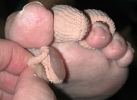
Any of your several types of nerves can be affected: your sensory nerves (which provide information about sensation, such as pain or heat), motor nerves (which carry signals to your muscles to provide tone and movement), or your autonomic nerves (which control reflexive, or non-voluntary body functions, such as breathing, heartbeats, and digestion). Well, kind of. Losing motor nerves in the foot may mean that the muscles in your feet will weaken. This weakening can cause deformities such as claw toe (toes that curl down into the soles of your shoes like bird claws), which can lead to serious discomfort as they rub against the inside of your shoe and form calluses. If coupled with sensory nerve damage, these calluses can become serious problems. Imagine you have an infected toenail. Now, imagine if you still had that infected toenail, but couldn't feel it. Or if you had a blister on the bottom of your foot and kept walking on it. Or a splinter. They look similar to Oribis.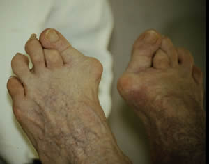
You may need to wear a surgical shoe with an open toe for several weeks following your operation. You may need to use crutches to help you avoid putting weight on your toes. This deformity occurs when the joint of the little toe becomes permanently contracted.
Sometimes a change in activity, shoes, or weight gain can make a bunion or hammertoe seem suddenly very painful. Orthotics, custom orthopedic foot supports, can help with mild hammer toes and bunions. Orthotics may slow down or perhaps prevent the progression of bunions and hammer toes.
Diabetic? You Could Be At Risk For Foot Ulcers
Keep your feet, elbows and other areas of your body free from diabetes skin problems with our selection of diabetes lotion. Understanding diabetic foot problems can be an important step (no pun intended) in learning to manage your diabetes and avoid the more serious complications. Armstrong et al. provide an overview of the evaluation and management of diabetic ulcers. Serious complications of diabetes can include foot ulcers. The pain started to ease off after taking your product for about six weeks, I will continue using your treatment as it is the first time in the four and a half years that I have felt so positive and hopeful. Many times, people aren't even aware that they have a wound on their feet, because neuropathy makes feet numb. Typically, diabetic foot infections are polymicrobial (more than one type of bacterium). In recent years, staph strains have evolved to be more resistant to antibiotics. The ulcers and open sores that can occur in diabetic feet can put you at risk for contracting MRSA in addition to other infections. MRSA is a type of staph infection that is resistant to many different antibiotics. One is called a "nosocomial" infection, meaning that it's an infection that is transmitted mostly in healthcare settings. Amato AA, et al. Peripheral neuropathy. Data on cellulitis in the diabetic lower limb and its outcome is lacking. The only known classifications for cellulitis and necrotizing fasciitis is Eron's classification7 and the one proposed by Wong et al.8 Eron's classification is a general classification of cellulitis and, as a whole, guides the admission and treatment modes. Stage 1 cellulitis has all the classical features of infection including redness, local warmth, and edema (Figure 1). No abscess or any localized skin necrosis is present. The first is that the necrotic tissue could have pus collection underneath which can cause spreading of the infection. This can result in a range of different problems. However, an infection this deep has access to the circulatory system and threatens to spread to other organs and body parts. One of the best ways to maintain your diabetic health is to control your diet and make better choices at mealtime. Without a doubt, your Foot Conditions Balm works like miracles. Diabetic patients need to wear shoes whenever they are outside.
 All consecutive eligible adult patients (age >18 years) with a diabetic foot lesion corresponding to Wagner 2 to 4 that has continuously existed for a minimum of four weeks, who meet all inclusion criteria and no exclusion criteria, may be included in the trial. Chronic diabetic foot wounds after adequate wound pretreatment (debridement and/or wound cleansing) as well as amputation wounds resulting from a planned amputation underneath the upper ankle joint (lat.: Articulatio talocruralis) may be considered for inclusion. If a sufficient covering is not possible, the patient should not be included in the study Patients may not be included if they are receiving anticoagulation therapy or are suffering from a higher grade impaired clotting function and have a heightened risk for bleeding with relevant circulatory effects if those patients are in outpatient care during the time of inclusion. Complete wound closure can be achieved either by surgery or by secondary intention. The skin is dry and itchy. DFCon is the premier international, interdisciplinary diabetic foot conference in North America. In its 13th year, the course is designed for the wide spectrum of generalists and specialists who diagnose and manage the diabetic foot. Didactic talks, panel discussions, Q&A sessions, specialty symposia and workshops will delve into diagnostic and interventional strategies for diabetic foot ulcers and amputation prevention. If you are interested in exhibiting at Diabetic Foot Global Conference 2014, please review the various levels and marketing opportunities still available. Valley Presbyterian Hospital's Amputation Prevention Center (APC) has a unique team-based approach to amputation prevention, involving patients as part of the care. The state-of-the-art 4,000 square foot facility is the only center of its kind on the West coast that solely focuses on preserving limbs. Moisturize your feet. Manage your diabetes. People who have diabetes should visit a doctor or podiatrist (foot doctor) regularly for preventative foot care and whenever a foot condition, such as an ulcer, ingrown toenail, plantar lesion or foot pain develops. Catching and treating foot problems early reduces the chances of an amputation. Keeping your blood glucose (sugar) in good control and taking care of your feet every day can help you avoid serious foot problems.